English
 Français
News
Home / News / Industry News / Alginate vs Hydrofiber Dressings: Hemostasis and Gel Formation Mechanisms

Your trusted medical supplies manufacturer.
No. 10 Ousheng Avenue, Pingwang Town, Wujiang District, Suzhou City, Jiangsu Province, China
+86 133 5801 3454
jasonzhangcas@gmail.com
Content
Exudate signals a healing wound, but it creates a practical engineering problem. A dressing must absorb fluid, keep the wound bed moist, survive removal in one piece and, when a wound is bleeding, help the body form a clot instead of washing it away. Alginate wound dressings and hydrofiber dressings solve that problem in the same broad way: both convert liquid into gel. Beyond that similarity, their chemistry, gel structure and hemostatic behaviour differ considerably.
This comparison focuses on the two questions that matter most in clinical evaluation and in product sourcing: how each dressing supports hemostasis, and how each one forms its gel.
A moist wound bed supports faster keratinocyte migration and granulation than a dry one, but the balance is narrow. Pooled exudate macerates the periwound skin and encourages bacterial growth, while a wound that dries out crusts over and stalls. Gelling fibrous dressings hold the middle ground, because the gel keeps fluid in contact with the wound bed under control instead of letting it spread sideways.
Both families are classed as high-absorbency, interactive dressings for wounds with moderate to heavy exudate, including cavity wounds. Both also rely on a secondary dressing, such as an absorbent pad, a foam or a film, to hold the gel in place and take up fluid that the primary dressing cannot.
Key point: both dressing families work by gelling, but only calcium alginate releases an ion that takes an active part in the clotting cascade.
Alginate fibres come from brown seaweed. Sodium alginate is extracted, spun into fibre and treated with a calcium salt so that the soluble polymer becomes insoluble calcium alginate. The result is a soft, highly conformable fleece supplied as flat pads, ropes and ribbons; the rope format makes alginate practical for packing narrow cavities and sinus tracts.
The hemostatic behaviour of calcium alginate follows from the material itself. When the fibre meets wound fluid, calcium held in the fibre lattice is released into the wound bed. Calcium is Factor IV in the coagulation cascade and acts as a cofactor in platelet activation and aggregation, in the conversion of prothrombin to thrombin, and in the polymerisation of fibrin into a stable clot.
Two secondary effects reinforce this. The fibre concentrates platelets and clotting factors at the wound surface as it takes up fluid, and the gel that forms holds the developing clot in place, a mild tamponade effect. That is why calcium alginate is a common choice for bleeding surfaces after debridement, for graft donor sites and for surgical wounds that ooze blood.
The effect has limits. Alginate dressings support the natural clotting process; they are not a substitute for surgical hemostasis in major arterial bleeding, and much of the supporting evidence is in vitro work and case series rather than large randomised trials.
Gelling is the same ion exchange running in reverse. Sodium ions in the exudate displace calcium from the fibre, and the fibre reverts to water-soluble sodium alginate, which swells into a soft hydrophilic gel. The gel keeps the wound bed moist while fluid is held inside the dressing structure.
Two consequences follow. Gelling needs moisture, so on a dry or lightly exuding wound the fibre stays intact and can adhere. And because the gel is relatively fluid and may spread, a secondary absorbent dressing plus monitoring of the wound margin are standard practice.
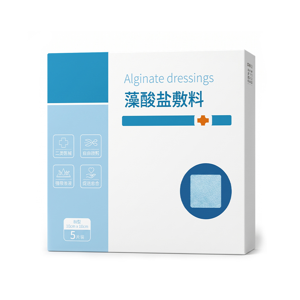
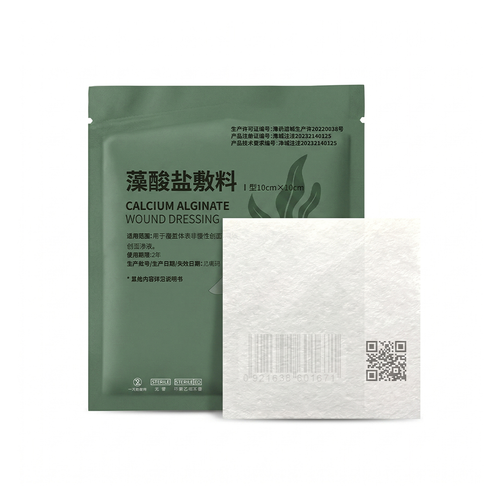
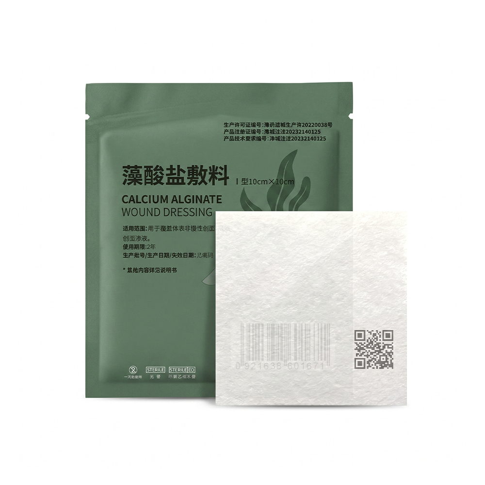 Sterile Calcium Alginate Wound Dressing 10 cm x 10 cmSterile calcium alginate dressing for moderate to heavy exudate; forms a soft non-adherent gel that supports moist healing and gentle removal.View Product →
Hydrofiber is a specific fibre technology rather than a generic description. The fibre is spun from sodium carboxymethylcellulose (CMC), a cellulose derivative that hydrates on contact with fluid. It is supplied as flat pads, cavity ribbons and bordered versions, and is also available with a silver component.
Gel formation here is hydration rather than ion exchange. Exudate contacts the CMC fibres, which swell and bind water into their structure, converting the dressing into a cohesive gel sheet that stays in one piece. Because fluid is locked inside the fibre network, the gel does not weep moisture back onto the periwound skin, and because the fibres wick fluid vertically rather than sideways, the risk of maceration at the wound margin is limited.
The cohesive gel also conforms closely to the wound bed, fills dead space in cavities and is normally removed in one piece, which makes residue checks straightforward and reduces discomfort at dressing changes.
Hydrofiber dressings contain no calcium, so they do not feed the clotting cascade the way calcium alginate does. Their contribution to bleeding control is mechanical: as the fibre takes up fluid, platelets and proteins at the wound surface are concentrated, and the gel layer covers the bleeding point and supports clot formation. Reports describe hydrofiber used successfully on oozing surgical wounds and donor sites, but the calcium-mediated pathway that defines alginate hemostasis is absent.
| Property | Alginate wound dressing | Hydrofiber dressing |
|---|---|---|
| Base polymer | Calcium alginate from brown seaweed | Sodium carboxymethylcellulose fibre |
| Trigger for gelling | Sodium-calcium ion exchange | Hydration and swelling of CMC fibres |
| Calcium release | Yes, calcium ions pass into the wound bed | None |
| Main hemostatic mechanism | Calcium-dependent support of the clotting cascade plus a gel barrier | Gel barrier and concentration of platelets and clotting factors |
| Gel character | Soft and hydrophilic, can be relatively fluid | Cohesive gel sheet, removed in one piece |
| Fluid handling | High absorbency, gel may spread laterally | Vertical wicking, fluid locked in the fibre |
| Cavity formats | Ropes and ribbons | Ribbons and pads |
| Dry wounds | Fibre can adhere if it does not gel | Needs moisture to gel, unsuitable when dry |
| Typical secondary dressing | Absorbent pad or foam | Foam or film, depending on exudate level |
Selection follows the wound rather than the brand, and a few rules cover most cases.
Moisture balance is the most common reason a gelling dressing underperforms. If a wound becomes too wet or dries out beneath the dressing, the cause is usually the dressing choice or the change interval rather than the wound itself. Signs that a dressing is leaving a wound too wet or too dry are worth checking before switching products.
Where a wound is no longer exuding heavily, and protection matters more than absorption, a hydrocolloid wafer often fits better than either gelling family.
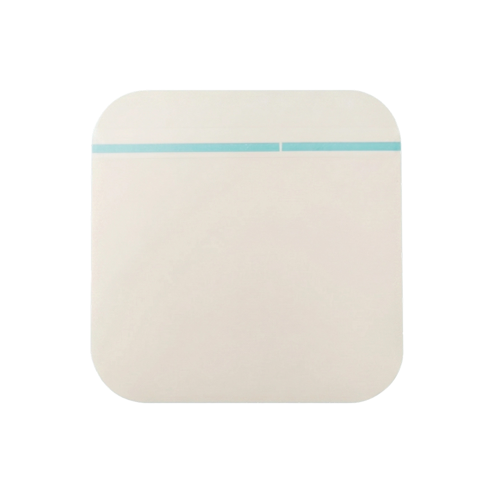 Hydrocolloid Wound Dressing 10x10 cm for Light ExudateHydrocolloid dressing for light-to-moderate exudate that creates a moist healing environment, provides a waterproof barrier, and can be cut to fit.View Product →
For distributors, first-aid suppliers and hospital procurement teams, alginate and hydrofiber products often sit in the same tender or shelf plan, so a supplier able to cover the whole gelling and foam segment has an advantage over one offering a single technology.
Suzhou Advan Medical Technology Co., Ltd. (ADVAN Medical), part of the ADVAN Group, has manufactured medical consumables and dressings since 2003 and exports to more than 60 countries. The company operates as a manufacturer and exporter with ODM and OEM services, works to ISO 13485:2016 with a documented inspection programme, states that most products carry CE marking, and describes its Suzhou plant as FDA compliant. Its wound care programme covers calcium alginate and alginate dressings in 10 cm x 10 cm formats alongside hydrocolloid, foam, silicone foam, silicone contact layer, NPWT sponge, transparent film and sterile wound dressing products, so a complete exudate-management range can be assembled from one source.
Buyers who need a specific size, format or private-label configuration can review the alginate wound dressing range and discuss specifications through the ODM and OEM service.
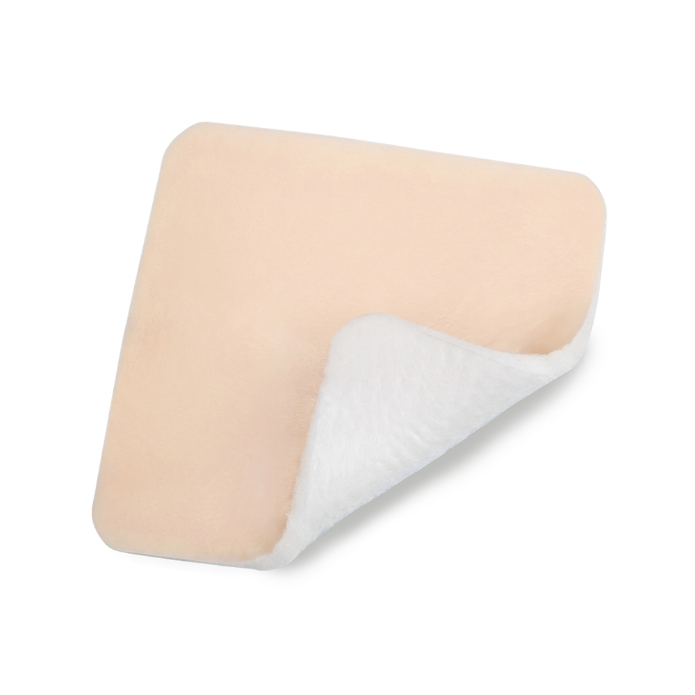 Borderless Silicone Foam Dressing for Moderate to Heavy ExudateBorderless silicone foam dressing absorbs excess exudate, maintains moist healing, and allows painless removal without tearing fragile tissue on moderate to heavy wounds.View Product →
Yes, within limits. Calcium alginate releases calcium ions into the wound bed, and calcium is a cofactor in the clotting cascade. The fibre also concentrates platelets and clotting factors, and the resulting gel holds the clot in place. This supports hemostasis on oozing or lightly bleeding surfaces, but it does not replace surgical hemostasis in major bleeding.
It can be used on wounds that ooze or bleed lightly, because the gel layer covers the bleeding point and helps concentrate clotting factors. However, hydrofiber contains no calcium, so its action is mechanical rather than biochemical. Where bleeding control is the primary aim, calcium alginate is normally the more direct choice.
Gelling depends on the sodium-calcium ion exchange, which requires fluid. On a dry or minimally exuding wound there is not enough exudate to convert the fibre, so the dressing stays intact and can adhere. A hydrogel can donate moisture under a gelling dressing, or a different dressing class can be selected.
Yes. Alginate and hydrofiber dressings are primary dressings. They are normally secured with an absorbent pad, a foam, a film or a bandage that keeps the gel in place and takes up any fluid the primary dressing cannot hold.
The two technologies look closer on a specification sheet than they behave at the bedside, and further apart in a wound than their appearance suggests. Calcium alginate earns its place where a wound bleeds as well as exudes, because calcium release links the dressing directly to the clotting cascade. Hydrofiber earns its place where the priority is a cohesive, vertically wicking gel that protects fragile skin and comes out in one piece. Most wound care ranges need both, and the choice between them is a clinical judgement about bleeding and exudate rather than a preference for one material.
Leading custom medical supplies manufacturers, including a full range of Wound Dressing, Hydrocolloid Medical Products.


Copyright © 2026 by Suzhou Advan Medical Technology Co., Ltd. All Rights Reserved. This website is intended for regions outside China. Approval No:20240008

 Previous